
Skin Lesions, Moles and Skin Cancers in Turkey
A skin lesion can range from a harmless mole or skin tag to a precancerous or malignant growth.
Not every bump, pigmented spot or rough patch on the skin is the same. Some lesions are completely benign, while others may require closer assessment, biopsy or surgical removal.
At Anka Plastic Surgery, skin lesions are evaluated according to their appearance, depth, location and clinical behaviour before deciding on the most appropriate treatment.
What Is a Skin Lesion?
A skin lesion is any area of skin that looks or feels different from the surrounding skin.
It may be:
- flat or raised,
- pigmented or skin-coloured,
- smooth or rough,
- stable for many years,
- or gradually changing over time.
Common skin lesions include moles, skin tags, seborrhoeic keratoses and actinic keratoses.
Some are benign, while others may represent or develop into skin cancer.
Moles and Dermal Naevi
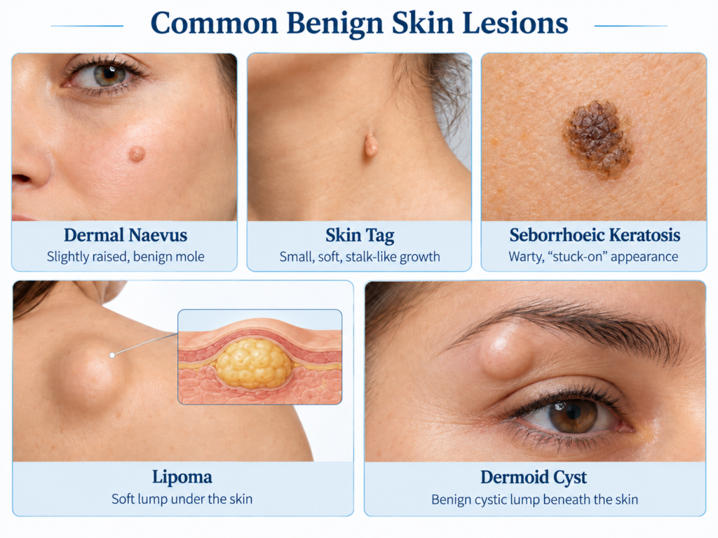
Moles, medically called naevi, are common skin lesions.
They may be flat, slightly raised or clearly elevated above the skin surface.
A dermal or intradermal naevus is usually a raised, skin-coloured or lightly pigmented mole that extends into the deeper layers of the skin.
For deeper lesions such as dermal naevi, surgical excision is often more appropriate than treating only the surface.
On the face, the incision is planned carefully according to natural skin lines and closed with fine sutures, with the aim of making the final scar as discreet as possible.
Skin Tags
Skin tags are small, soft, often stalk-like growths that commonly occur around the neck, eyelids, underarms and skin folds.
They are benign and are different from true melanocytic moles.
Small superficial skin tags can often be treated with laser or other minor procedures.
Seborrhoeic Keratosis
Seborrhoeic keratoses are common benign skin growths that become more frequent with age.
They may appear brown, grey or skin-coloured and often have a slightly raised, rough or “stuck-on” appearance.
Most do not require medical treatment.
Removal may be considered if they:
- become irritated,
- catch on clothing,
- cause cosmetic concern,
- or have an appearance that makes the diagnosis uncertain.
Selected superficial lesions may be suitable for laser or other surface treatments.
Actinic Keratosis
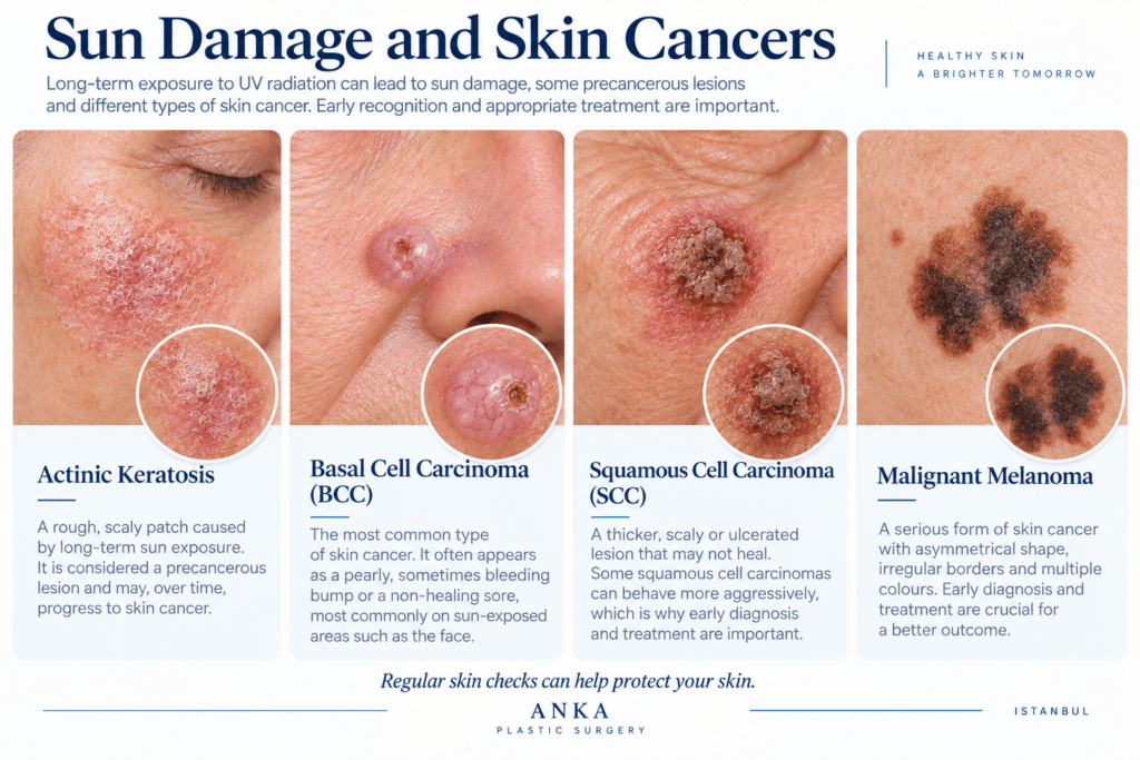
Actinic keratosis is associated with long-term sun damage.
It is most commonly seen on areas such as:
- the face,
- nose,
- ears,
- forehead,
- scalp,
- and backs of the hands.
These lesions often appear as dry, rough or scaly patches.
Actinic keratosis is important because it is considered a precancerous lesion, and a proportion can progress to squamous cell carcinoma.
For this reason, persistent rough or changing lesions on sun-exposed skin should be assessed rather than treated simply as cosmetic spots.
Lipoma
A lipoma is a benign growth made of fatty tissue. It usually presents as a soft, mobile lump under the skin and tends to grow slowly over time.
Lipomas are commonly found on the:
- neck,
- shoulders,
- back,
- upper arms,
- trunk,
- and thighs.
Most lipomas are harmless and do not require treatment unless they become painful, increase in size, cause pressure symptoms or create a cosmetic concern.
When removal is indicated, the usual treatment is surgical excision, with the aim of removing the fatty mass together with its capsule when possible.
Very large, rapidly growing, painful or unusually firm masses should be assessed carefully before treatment, as not every soft-tissue lump is necessarily a simple lipoma.
Dermoid Cyst
A dermoid cyst is a benign congenital cystic lesion that develops beneath the skin.
It may contain skin cells, sebaceous material and, in some cases, hair-bearing structures.
Dermoid cysts are commonly found around the:
- eyebrow,
- forehead,
- eye area,
- scalp,
- and neck.
They usually grow slowly and may remain painless for many years. Over time, however, they can become more noticeable, inflamed or cosmetically bothersome.
Treatment is usually surgical excision. The aim is not simply to drain the cyst, but to remove it together with its wall whenever possible to reduce the risk of recurrence.
For dermoid cysts located near the eye, eyebrow or deeper facial structures, preoperative imaging may sometimes be required to assess their relationship with the surrounding anatomy.
When Should a Skin Lesion Be Checked?
Most skin lesions are benign, but some changes should prompt medical assessment.
These include:
- rapid growth,
- change in colour,
- irregular borders,
- repeated bleeding,
- ulceration,
- persistent crusting,
- a sore that does not heal,
- or a lesion that looks noticeably different from the others.
In these situations, diagnosis should come before cosmetic treatment.
Skin Cancers
Basal Cell Carcinoma (BCC)
Basal cell carcinoma is one of the most common forms of skin cancer.
It is particularly common on sun-exposed areas of the face, including the nose, cheeks, forehead, ears and around the eyes.
BCC usually grows slowly and rarely spreads to distant organs. However, if left untreated, it can progressively invade surrounding tissues.
Treatment is therefore important even when the lesion appears small or slow-growing.
The treatment plan depends on factors such as the size, location and pathological characteristics of the tumour.
Squamous Cell Carcinoma (SCC)
Squamous cell carcinoma is another important skin cancer and may also be related to chronic sun exposure.
Some SCCs develop from areas of actinic keratosis.
Compared with BCC, certain SCCs may behave more aggressively and have a greater potential to spread to lymph nodes or other areas.
Treatment depends on the size, depth, location and pathological features of the tumour.
Malignant Melanoma
Malignant melanoma develops from melanocytes, the pigment-producing cells of the skin.
It may arise within an existing mole or appear as a new pigmented lesion.
Early diagnosis is particularly important.
One practical way to assess suspicious moles is the ABCDE rule:
- A – Asymmetry: one half looks different from the other
- B – Border: irregular or poorly defined edges
- C – Colour: different colours or shades within the same lesion
- D – Diameter: increasing size may be a warning sign
- E – Evolution: any significant change over time
Bleeding, ulceration or a mole that looks very different from the others should also be assessed.
Should a Suspicious Mole Be Removed with Laser?
If a pigmented lesion is clinically suspicious or the diagnosis is uncertain, laser treatment is generally not the first choice.
Destroying a lesion with laser may remove tissue that could otherwise be examined under the microscope.
In these situations, surgical removal and histopathological examination may be more appropriate.
How Are Skin Lesions Removed?
The treatment depends on the type, depth and clinical features of the lesion.
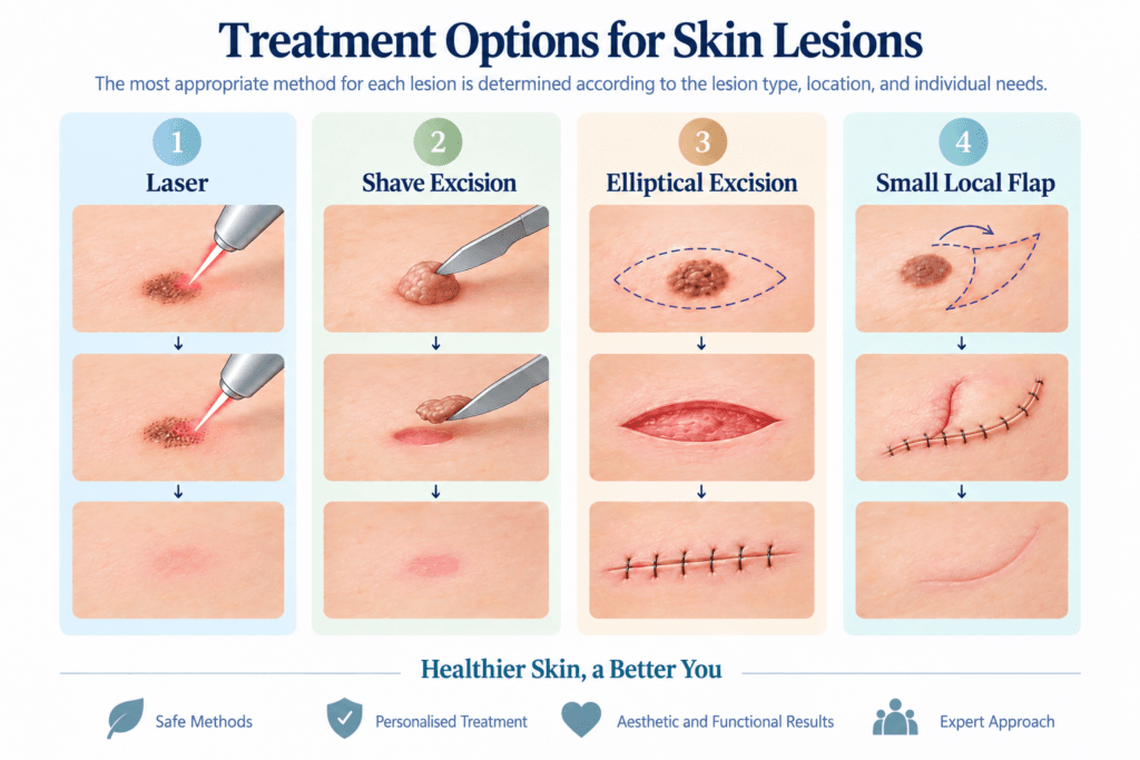
Surgical Excision
Surgical excision may be used for dermal naevi, suspicious lesions and lesions that require pathological examination.
Micro-Excision
Very small, selected facial moles may sometimes be removed through a tiny opening and closed with a single fine stitch.
Laser Treatment
Laser may be appropriate for selected superficial benign lesions such as skin tags and some seborrhoeic keratoses.
No single treatment method is suitable for every lesion.
Skin Cancer Surgery and Facial Reconstruction
When a skin cancer is removed from the face, treatment does not always end with tumour excision.
The resulting defect may also require reconstruction.
Depending on its size and location, reconstruction may involve:
- direct closure,
- local flaps,
- skin grafts,
- or, in more complex defects, cartilage grafts and structural reconstruction.
This is particularly important in areas such as the nose, lips, eyelids and ears, where both appearance and function need to be preserved.
The aim is not only to remove the lesion appropriately, but also to reconstruct the resulting tissue loss in a way that respects the anatomy and function of the area.
Conclusion
Not every skin lesion requires the same treatment.
Some benign superficial lesions may be suitable for laser, while deeper, changing or suspicious lesions may require surgical excision and histopathological examination.
In facial skin cancer surgery, treatment may also involve reconstruction in order to preserve both appearance and function.
For patients with a mole, changing skin lesion or facial skin tumour, an initial assessment can help determine whether observation, minor treatment, surgical excision or a more detailed reconstructive plan is appropriate.